
Normal values of regional longitudinal systolic strain obtained by

Value of Speckle Tracking–Based Deformation Analysis in Screening Relatives of Patients With Asymptomatic Dilated Cardiomyopathy
Reference values for two-dimensional myocardial strain echocardiography of the left ventricle in healthy children, Cardiology in the Young
Reference values for two-dimensional myocardial strain echocardiography of the left ventricle in healthy children, Cardiology in the Young
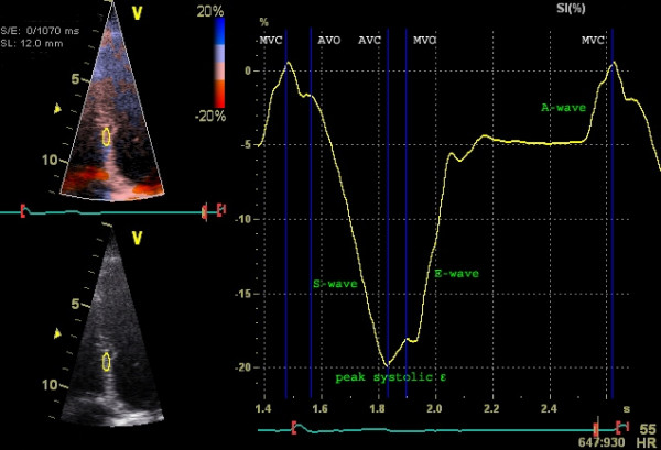
Strain, strain rate and speckle tracking: Myocardial deformation – ECG & ECHO

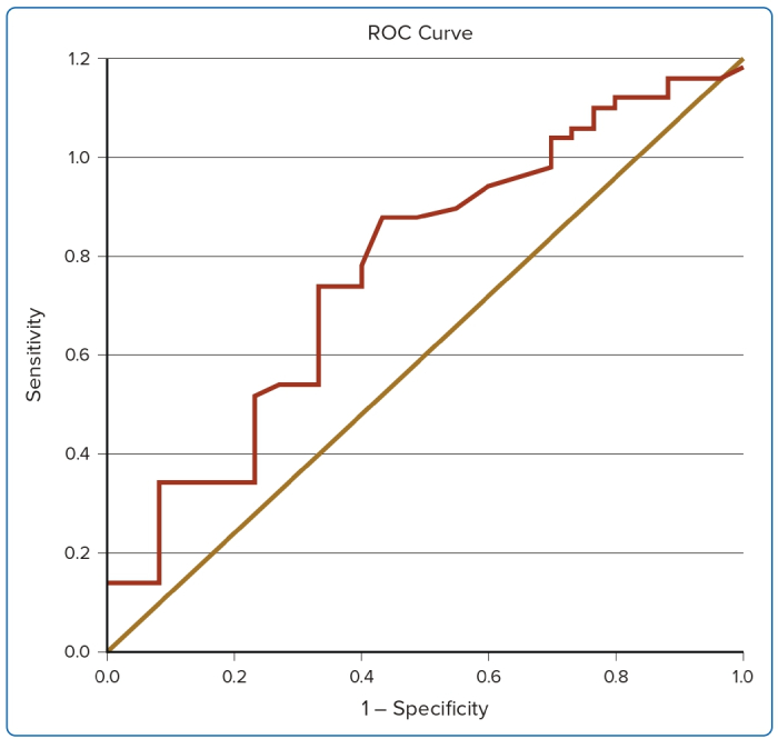
Novel regional longitudinal strain by speckle tracking to detect significant coronary artery disease in patients admitted to the emergency department for chest pain suggestive of acute coronary syndrome

2- and 3-Dimensional Myocardial Strain in Cardiac Health and Disease

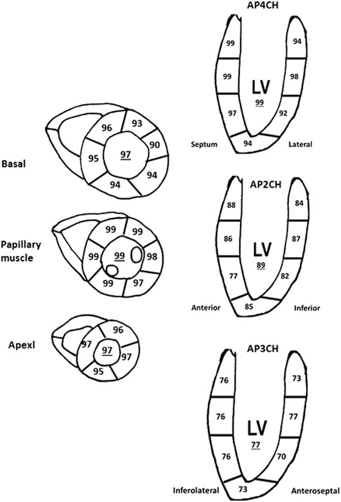
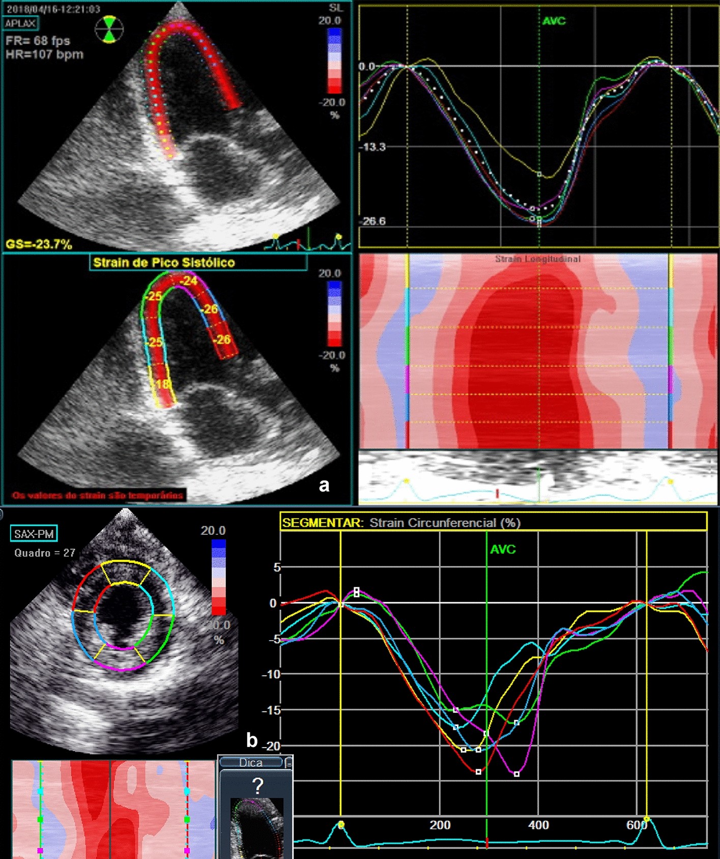
Bulls-eye figure of peak systolic strain values in the left ventricle
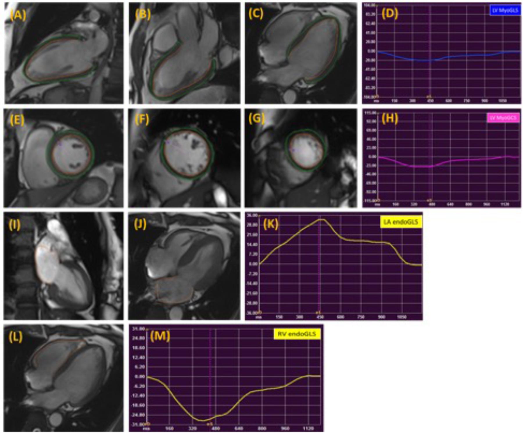
Frontiers Normal Values of Myocardial Deformation Assessed by Cardiovascular Magnetic Resonance Feature Tracking in a Healthy Chinese Population: A Multicenter Study

Longitudinal strain per level: apex, mid, and basal. Left ventricular
Evaluation of ventricular systolic function by speckle tracking technique in patients with biliary atresia before and after liver transplantation
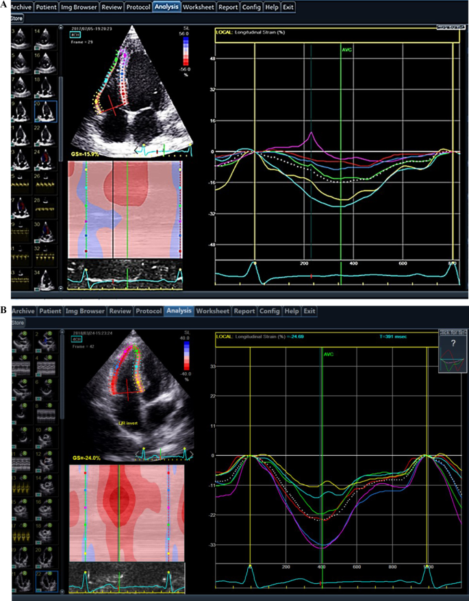
Evaluation of Subclinical LV Systolic Dysfunction by GLS Using 2D-STE

Myocardial Deformation in the Pediatric Age Group: Normal Values for Strain and Strain Rate Using 2D Magnetic Resonance Feature Tracking - Voges - 2022 - Journal of Magnetic Resonance Imaging - Wiley Online Library
Apical but not basal RV strain reflects right ventricular dysfunction in patients with dilated cardiomyopathy, Egyptian Journal of Radiology and Nuclear Medicine

Average peak systolic longitudinal strain values of each left

Principles and Practical Aspects of Strain Echocardiography
Normal values of regional longitudinal systolic strain obtained by
- Best Price $ 81.50. Good quality and value when compared to berghoff.ir similar items.
- Seller - 768+ items sold. Top-Rated Plus! Top-Rated Seller, 30-day return policy, ships in 1 business day with tracking.
People Also Loved
-

LV x YK Christopher Backpack Monogram Taurillon Leather - Men - Bags
Buy It Now 15d 20h -

Avenue Slingbag NM Monogram Macassar - Bags
Buy It Now 21d 7h -

Louis Vuitton Rose De Vents EDP 3.4oz 80% Full Tester- Batch Code 1L01
Buy It Now 9d 16h -

Louis Vuitton Brown Monogram Canvas Bi-Fold Small Compact Wallet
Buy It Now 4d 13h -

Louis Vuitton Duomo Damier Ebene Handbag Satchel Women Brown Purse Tote LV Gold
Buy It Now 8d 12h -

Officine Universelle Buly 1803
Buy It Now 17d 5h -

Victoria Beckham Chain Pouch With Strap Khaki Leather Shoulder Bag
Buy It Now 2d 23h -

PHOTOS: 2023 Las Vegas Helldorado Days Parade – Part Two – AmericaJR
Buy It Now 13d 5h -

Pharrell - Luis Vuitton dinner jacket. Fashion trends winter, Trendy prom suits, Fashion
Buy It Now 11d 15h -
Louis vuitton buy and sell Philippines
Buy It Now 6d 20h -

Dooney & Bourke Handbag, Nylon North South Triple Zip Crossbody
Buy It Now 5d 15h -

Veronica Hoop Earrings in Gold
Buy It Now 15d 21h -
 by 碧aoi")
Shop Louis Vuitton Attitude Sunglasses (Z0259U) by 碧aoi
Buy It Now 26d 5h -

Cheval Blanc St-Barth Isle de France
Buy It Now 26d 11h -
 – Kool Breeze Solar Hats")
Kool Breeze Solar Men's Cowboy Straw Hat (Band) – Kool Breeze Solar Hats
Buy It Now 12d 16h -

🌞MICHAEL KORS HAMILTON LARGE NS LUGGAGE BROWN SAFFIANO LEATHER TOTE BAG🌺NWT!
Buy It Now 7d 11h -

Louis Vuitton Supreme Camouflage Canvas Jacket
Buy It Now 8d 18h -

The Season's Most Covetable Accessories? Boots—Shiny, Sparkly
Buy It Now 11d 5h -
ZooZatz Louisville Cardinals Scarf and Cuffed Knit Hat with Pom Set
Buy It Now 25d 7h -

Louis Vuitton Empreinte - Embossed Leather Handbags, Wallets
Buy It Now 10d 7h -

Louis Vuitton Emilie wallet damier azur with rose ballerine interior
Buy It Now 16d 10h -

Louis Vuitton x NBA multiple wallet Review! A NBA rookie champ at
Buy It Now 17d 13h -

Nike Air Force 1 Low x LOUIS VUITTON LV 'White' 1A9V87 - KICKS CREW
Buy It Now 8d 16h -

LOUIS VUITTON LV CURVI FOLD iPhone 12 Mini Case Cover
Buy It Now 28d 16h

